Rotation Sensitivity
Minor patient rotation during acquisition may significantly alter imaging interpretation accuracy.
Advanced positioning workflows for craniocervical instability evaluation using precision imaging alignment and AI-supported positioning technology.
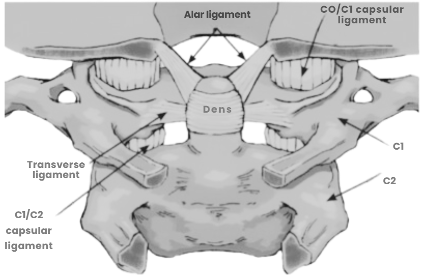
“The Cervicocranium is defined as extending from the skull base through the second cervical intervertebral disk. Of the entire cervical spine, the Cervicocranium is the most difficult to evaluate radiographically because of its complex anatomy, the physiologic motions and normal variants that may simulate acute injury, and the inherent difficulty in obtaining a diagnostic study of the Cervicocranium in both frontal and lateral projections. These factors have particularly adverse implications in patients clinically suspected of having blunt Cervicocranial trauma, when it is impossible to obtain a useful open mouth view and given the fact that any injuries to the Cervicocranium are radiographically subtle.” Cervicocranium: its Radiographic Assessment." John H. Harris Jr, MD, DSc, Radiology 2001;218:337-351, P1
“AP OM neck lateral flexion weight bearing X-ray imaging study is considered a gold standard diagnostic test for determining CCI in the sagittal plane.” Occult Hypermobility of the Craniocervical Junction: A Case Report and Review”. Page 444 | june 2011 | volume 41 | number 6 | journal of orthopaedic & sports physical therapy
Proper evaluation of the craniocervical junction (CCJ) requires pure orthogonal alignment during physiologic weight-bearing positioning.
The atlanto-axial joint is a highly rotational structure responsible for nearly 70 percent of total neck rotation.
Even minor rotational deviation during imaging acquisition can create false positive imaging findings and unreliable diagnostic interpretation.
Minor patient rotation during acquisition may significantly alter imaging interpretation accuracy.
Proper 15-degree cephalad tilt positioning introduces additional workflow and alignment complexity.
Different imaging geometry angles between X-ray and positioning systems require additional precision adjustment.
Patient movement during CT or MRI acquisition may produce non-diagnostic imaging results.
Coupling Smartray® GLX 0.1 with stationary X-ray imaging workflows improves positioning reproducibility and supports interrater reliable evaluation of craniocervical instability.
Conventional MRI, CT, or uncontrolled X-ray positioning may have reliability limitations due to patient motion and positioning inconsistency.